
About us
The Cell Analysis Core provides researchers access to state-of-the-art instrumentation in flow cytometry, confocal microscopy, and consultative scientific services to support their research goals. Our facility provides comprehensive training in cross-translational cytometry and imaging, including experimental design, sample preparation, instrument operation, and data analysis.
Our mission
The mission of the Cell Analysis Core is to provide centralized services, training, access, and support to researchers in the use of cutting-edge technology for the study and manipulation of biological samples using flow cytometry and confocal microscopy equipment.
We are also striving to establish an active training and consultation program that supports the use of cytomics and laser scanning microscopy, improving not only research productivity but also the competitiveness of students and fellows trained on our campus.
Core information
- HoursMonday–Friday
8 a.m.–5 p.m.For after-hours use, contact us. - LocationBSE 3.108-1
- Phone(210) 458-4093
- Emailcell.analysis@utsa.edu
Instrumentation
-

Cell sorting
FACSAria II Cell Sorter
Equipped with four lasers, this instrument can detect 11 colors in a configuration similar to the LSR-II. To support research using BSL-2 human samples and samples that carry known pathogens or replication-defective retroviruses, the core provides sorting and cell analysis under a biosafety cabinet.
Configuration (PDF) -
Spectral flow cytometry
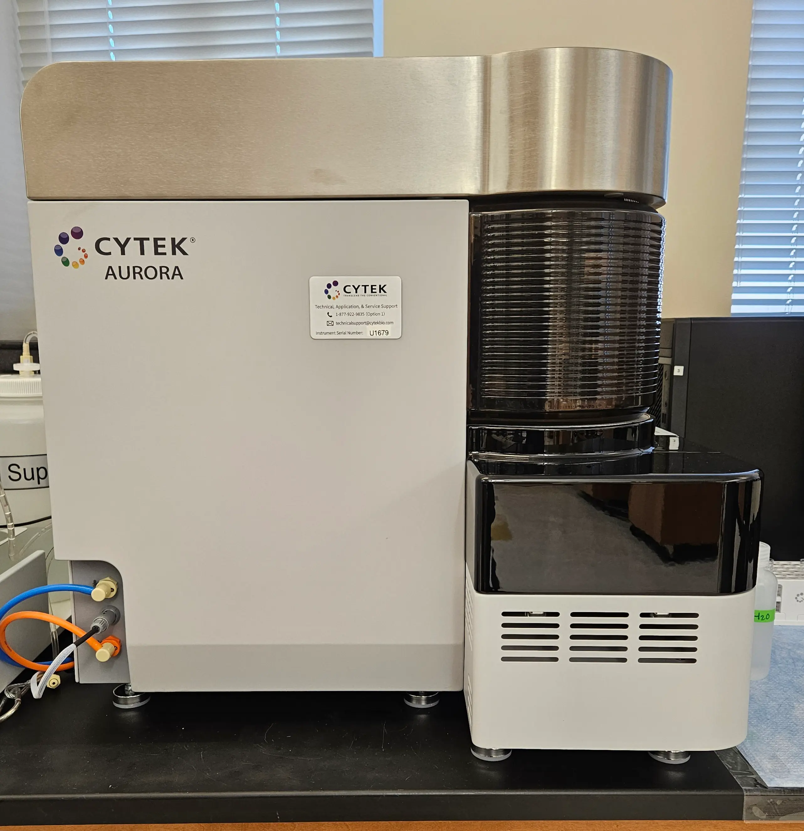
Cytek Aurora Spectral Cell Analyzer
This full-spectrum cell analyzer can detect up to 40 colors. It offers improved sensitivity and flexibility in panel design, from low- to high-complexity applications.
Lasers
- 355 nm
- 405 nm
- 488 nm
- 561 nm
- 640 nm
Detection
- 64 fluorescence channels
- Forward scatter (FSC)
- Blue laser side scatter (SSC)
- Violet laser side scatter (SSC)
-

Imaging flow cytometry
ImageStream Mk II Imaging Flow Cytometer
This four-laser, 12-channel imaging flow cytometer provides the capabilities of a regular cytometer and adds the ability to visualize individual cells microscopically at the same time. It captures detailed multispectral images of cells at rates of up to 5,000 cells per minute using 10 fluorescent channels and two brightfield channels.
Configuration (PDF) -

Live cell imaging
Zeiss LSM 710 NLO Two-Photon System
Includes an incubation chamber for live cell imaging.
Laser lines
- Diode 405 nm
- Diode 561 nm
- Tunable argon 458/477/488/514 nm
- Helium-neon 561 nm
- Chameleon Ultra II Ti:sapphire
Objectives
- 5X Plan-Apochromat/0.16 NA with DIC
- 10X Plan-Neo/0.3 NA with DIC
- 20X Plan-Apo/0.8 NA with DIC
- 40X Plan-Neo/1.3 NA oil with DIC
- 40X C-Apochromat/1.2 water
- 63X Plan-Apo/1.4 NA oil with DIC
Objectives are for the inverted Observer Z1 widefield fluorescence microscope.
-
Multiplex assays
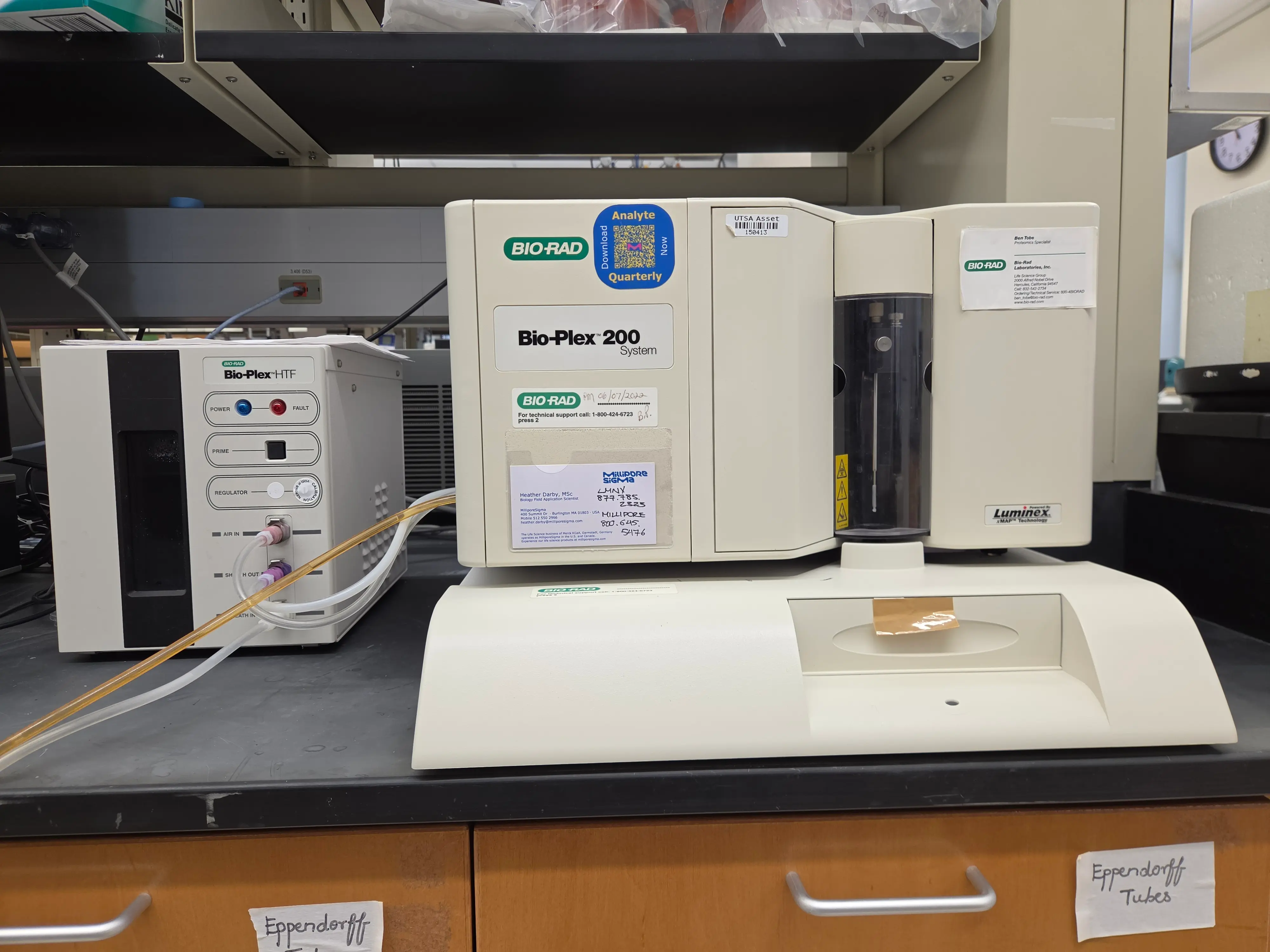
Bio-Plex 200
The Bio-Plex 200 offers high-degree multiplexing that dramatically increases the amount of useful information from rare or volume-limited samples, such as mouse and rat serum. It can analyze more than 9,600 assay points in 30 minutes in a 96-well plate by distinguishing up to 100 different families of color-coded, monodisperse polystyrene beads, each bearing a different homogeneous capture assay, in a single 50 µL sample.
Contact us
-
Sandra M. Cardona, Ph.D.
Research Core Director
Assistant Professor of Research -
Astrid E. Cardona, Ph.D.
Scientific Advisory Board Chair
-
Jose Caso, M.S.
Research Science Associate II
